
Cell function is determined by how DNA is expressed into proteins. That process includes two main steps — transcription, when messenger RNA (mRNA) makes copies of active genes, and translation, when mRNA guides protein assembly.

Knowing which genes are active at any given moment would make it possible to track the body’s response to factors in the environment or medication or to track the progression of an illness, along with tracking other key responses. Bioengineers at Rice University have developed a way to map transcription profiles in living brain tissue with great accuracy based solely on a blood sample.
“This is the first demonstration of measuring transcription for targeted genes nondestructively in living tissue,” said Jerzy Szablowski, assistant professor of bioengineering at Rice and the corresponding author on a study published in Nature Communications. “That means that we can actually select which gene we want to study and then see how it expresses over time within the same organism. That allows us to see what happens before, for example, a disease occurs and how gene expression changes as that disease progresses.”
The transcription-tracking tool combines two cutting-edge technologies ⎯ engineered reporter molecules called Released Markers of Activity (RMAs) pioneered by Szablowski and his team, and sensors that detect the presence of target mRNA in the cell then trigger the production and release of RMAs into the bloodstream.
“I combined two recent technologies to establish a new in vivo interface — monitoring of transcription in living tissues,” said Sho Watanabe, a postdoctoral researcher in the Szablowski lab who is a first author on the study.
Szablowski said the new method, called In-vivo Tracking of Active Transcription, or INTACT, is scalable and “in theory, should allow monitoring any gene by simply including its sequence in a genetic construct.” For instance, INTACT could be used to track the expression of genes associated with Parkinson’s, Alzheimer’s or even ones associated with a specific neural circuit.
“You do not have to make a bespoke reagent for each and every one these genes ⎯ instead, the targeting of genes is programmable,” Szablowski said.
The new method unlocks capabilities similar to next generation sequencing (NGS) and quantitative polymerase chain reaction (qPCR), impactful technologies that have allowed researchers to move from analyzing genes and molecules in a sample individually to tracking them collectively. However, NGS and qPCR require the destruction of analyzed samples, which means they can only be deployed in excised tissue or cells grown in a petri dish. In contrast, INTACT tracks gene expression in living tissue over time.
“In the future, we want to make this omics revolution possible in living tissue” Szablowski said. “This is the first step, and we are already working on the next ones.”
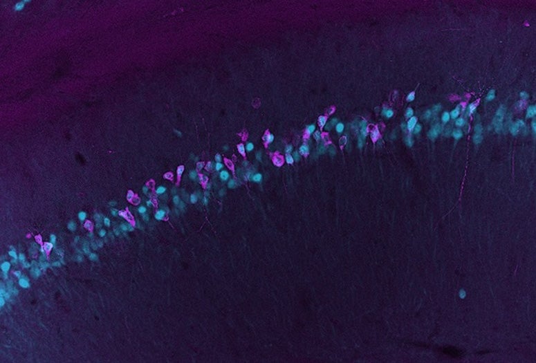
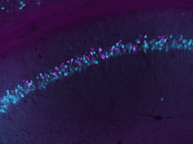
The researchers demonstrated the platform in an animal model, showing it could track three different brain regions at once. Szablowski said future development of the INTACT platform could lead to “highly multiplexed monitoring,” where large numbers of different genes, neural circuits or brain regions could be tracked simultaneously.

Watanabe added that he expects INTACT “could be applied for monitoring gene expression, not only in the brain but in other tissues as well.”
Before his work on RMAs as part of Szablowski’s team, Watanabe studied muscle biology with a focus on extracellular vesicles, molecules cells use to communicate with each other. His next project will draw on both areas of expertise to explore the use of synthetic mechanisms to enable communication between different organs and areas in the body. He said the three years he spent working on INTACT were an important stage in his professional development.
The research was supported by the David and Lucile Packard Foundation (2021-73005), the National Institute of Biomedical Imaging and Bioengineering (DP2EB035905), the Japan Society for the Promotion of Science postdoctoral fellowship and the U.S. National Science Foundation (1842494). The content herein is solely the responsibility of the authors and does not necessarily represent the official views of the funding organizations and institutions.
- Peer-reviewed paper:
-
Monitoring in vivo transcription with synthetic serum markers | Nature Communications | DOI: 10.1038/s41467-026-73486-2
Authors: Sho Watanabe, Sangsin Lee, Manwal Harb, Shirin Nouraein, Emma Raisley, Honghao Li, Nicolas Buitrago, Beatrice Pforr and Jerzy O. Szablowski
https://doi.org/10.1038/s41467-026-73486-2 - Access associated media files:
-
https://rice.photoshelter.com/galleries/C0000ozmLFGQBqew/G00005PPHha8n3yE/260522-Jerzy-Szablowski_Jones (Photos by Jared Jones/Rice University)