
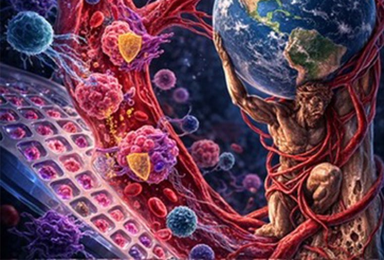
Metastasis, the spread of cancer from a primary tumor to other parts of the body, is difficult to study in the lab, in part because researchers lack reliable ways to recreate the conditions cancer cells encounter as they travel through the bloodstream.
Rice University bioengineers report a new platform designed to streamline one of the major challenges for metastasis research: Called the Advanced Tumor Landscape Analysis System, or ATLAS, the platform makes it easier to generate large quantities of cancer cell clusters that accurately model those involved in metastasis. Using the platform, the Rice team gained new insights into the mechanisms that enable cancer clusters to survive in the bloodstream during the metastatic process.
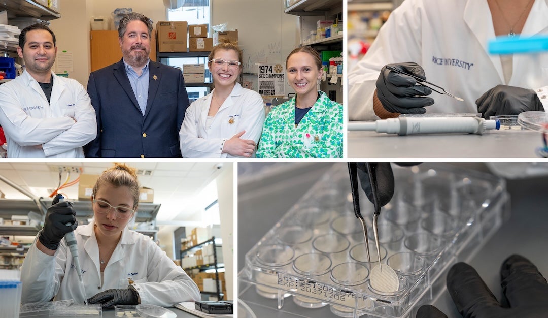
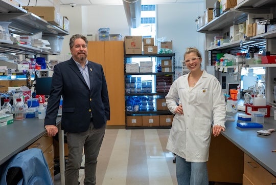
Developed in the lab of Michael King, Rice’s E.D. Butcher Professor of Bioengineering, ATLAS builds on earlier work using superhydrophobic surfaces, i.e. materials that strongly repel water. When droplets containing cells are placed on these surfaces, they bead up rather than spread out, encouraging cells to stick to each other and produce three-dimensional clusters.
“Metastasis is still poorly understood because adequate laboratory techniques to recreate this complex process are lacking,” said King, a Cancer Prevention and Research Institute of Texas Scholar who also serves as special adviser to the provost on life science collaborations with the Texas Medical Center.
The King lab has been working for years on new high-throughput approaches of creating clusters of cancer cells, both on their own and alongside stromal cells, which are noncancerous but are frequently found in the tumor microenvironment. To study what happens during the metastatic process, the researchers expose these models to conditions that closely resemble those in the body ⎯ either via animal models or laboratory blood flow experiments.
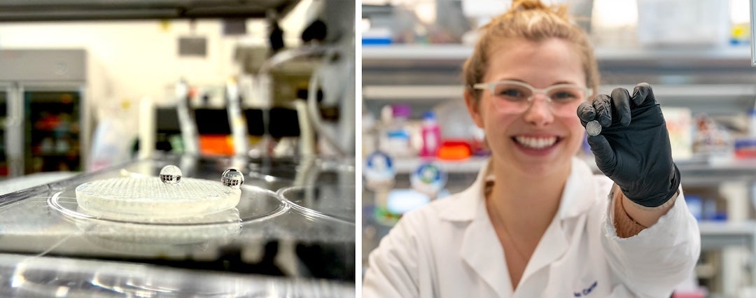
Compared to earlier methods, ATLAS takes less time to deploy and costs less to produce. It uses 3D-printed microwell arrays that are treated to create the same kind of water-repelling effect seen in nature, such as on a lotus leaf.
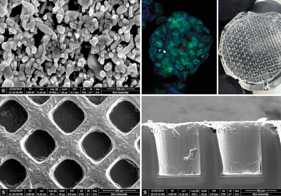
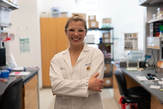
“The way this is achieved, both in nature and in the laboratory, is to create a surface that is rough on a nanoscale level, and then to coat the nanoscale bumps with a nonwetting substance such as Teflon or wax,” said Alexandria Carter, a doctoral student in the King lab who is the first author on the study. “Here, we achieved this for the first time through 3D printing, which means the method is scalable and easily adoptable by other labs.”
Going beyond method development into actual testing, the researchers used ATLAS to create clusters of prostate cancer cells, including ones containing a type of stromal cell called cancer-associated fibroblasts, or CAFs. Testing revealed that cancer clusters are more likely to survive when traveling in groups, especially when CAFs are present. These support cells actively help cancer cells withstand the stresses of circulation and continue to grow.
“One of the most exciting elements of our paper is that it does not just report on a new experimental method for other researchers to use, but it also reports new fundamental biological results,” Carter said. “Perhaps in the future the next generation of prostate cancer drugs will target these CAF ‘escorts’ as a way to prevent metastasis.”
Carter recently completed the Rice Innovation Fellows program and is working on establishing a startup company called Bionostic to commercialize ATLAS. Run by Rice’s Liu Idea Lab for Innovation and Entrepreneurship (Lilie), the program trains doctoral students and postdoctoral researchers to translate their research into breakthrough solutions for real-world problems.
Kyle Judah, Lilie executive director, said “a pre-requisite for bringing research beyond the bench is to be deeply passionate about the problem space, and Carter is the perfect example of an exceptionally driven and committed engineer willing this idea into reality.”
Models that are both realistic and practical make possible research that would otherwise take longer and be costlier to undertake.
“ATLAS makes it easier to study one of the most dangerous aspects of cancer,” King said.
- Peer-reviewed paper:
-
A Superhydrophobic 3D Cell Culture System Reveals the Mechanobiological Role of Cancer-Associated Fibroblasts in Prostate Cancer Metastasis | Advanced Healthcare Materials | DOI: 10.1002/adhm.202600011
Authors: Alexandria Carter, Abigail Fabiano, Ehsan Aalaei, Jason Deng, Dakota Rostant and Michael King
https://doi.org/10.1002/adhm.202600011 - Access associated media files:
-
https://rice.box.com/s/0w66n363y9p0vb2b3t1sjkpjru5bq41k
Photos by Jared Jones/Rice University
Microscopy images courtesy of Alex Carter/Rice University
B-roll by Brandon Martin/Rice University