

Rice University and collaborators in Switzerland have developed a new platform to grow human-derived nerve cells with Schwann cells, or supporting cells that form a protective coating called myelin around nerve fibers. This platform enables functional myelin in a three-dimensional lab-grown environment. Researchers confirmed that it worked by measuring an increase in the speed of electrical signals traveling across networks of connected nerve cells.

Developed at Rice and ETH Zurich, the platform combines human-derived cells, tissue-like materials and electrical measurements in one system. Because nerves contain many cell types and can be difficult to access without damage, researchers can use the model to study how myelin develops, how injury or exposure to toxins affects it and whether drugs or electrical stimulation can prevent its loss or promote repair. The platform can also be adapted to model the brain and measure complex cellular processes. The study was published Aug. 26 in Advanced Healthcare Materials.
“While the formation of myelin is exciting to see, we are even more excited that it is functional and changes how the nerve communicates,” said Christina Tringides, corresponding author and assistant professor of materials science and nanoengineering at Rice.
Creating a better home for nerve cells
Conventional surfaces used to grow cells in the lab, known as tissue culture plastic, make cells easy to visualize but are much more rigid than biological tissues such as the brain. As a result, cells do not receive the same physical cues they experience inside the body, Tringides said.
The researchers instead grew human-derived sensory neurons in hydrogels, soft, water-rich materials whose stiffness can be adjusted to more closely match nerve tissue. The neurons survived for more than 100 days.
Researchers grew the neurons alongside Schwann cells, which support healthy nerve function in the peripheral nervous system and form myelin by wrapping around axons, the long fibers that carry nerve signals.
“The environment matters to a cell,” Tringides said. “When it feels more like the body, we can see behaviors that are not possible to reproduce on plastic surfaces. Hydrogels let us recreate biological environments and support cells to carry out their native behavior.”
Seeing myelin and testing its function
The researchers combined the hydrogel with small channels that guide cell growth and placed the system on a high-density multielectrode array from Maxwell Biosystems, a device with many small electrodes that record neuronal activity in real time.
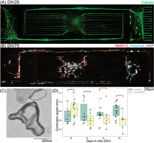
Called ‘hydroMEA,’ the platform lets the team control how cells are arranged while still providing a three-dimensional environment for growth. Imaging confirmed that myelin had formed around nerve fibers. Electrical measurements then showed that signals traveled faster when neurons were grown with Schwann cells, indicating the myelin was functional.
“That distinction is important,” Tringides said. “A microscope shows us what nerves look like. Measuring electrical activity tells us about nerve function. We were also very excited to see that the conduction velocities we measured were in the same range as those for human sensory nerves.”
Studying nerve damage and repair
With the platform in place, researchers can examine how nerve trauma or exposure to toxins affects neurons and myelin while tracking changes in electrical signaling.
The system also provides a way to test drugs or electrical stimulation for their ability to prevent demyelination, the loss or damage of myelin, or promote remyelination, the process of rebuilding it after damage.
“The next question is what happens when myelin is damaged and whether we can help it recover,” Tringides said. “We can use our hydroMEA platform to ask these questions, induce demyelination in the networks and study how drugs or electrical stimulation could promote remyelination.”
Other authors include Rice’s Nicole Baalbaki, Max Paxtian and Jeeho Sim; Blandine Clément, Cédric Pfister, Timothy Kurer, Julian Hengsteler, Sean Weaver, Tobias Ruff and János Vörös of the Institute for Biomedical Engineering at ETH Zürich; Céline Labouesse, Dhanajay Deshmukh, Lorenza Paganella and Mark Tibbitt of the Macromolecular Engineering Laboratory at ETH Zürich; and Julia Lehmann, Vilius Dranseika and Lukas Sommer of the University of Zürich.
The study was supported by an ETH Zurich Postdoctoral Fellowship, ETH Zurich, the Swiss National Science Foundation and Rice.