
Rice University bioengineer Julea Vlassakis has won $1.1 million in federal funding for a project researching Ewing sarcoma, an aggressive bone and soft tissue cancer that mainly affects children, teenagers and young adults. Vlassakis is one of 10 researchers to obtain a Peer Reviewed Cancer Research Program Career Development Award as part of the U.S. Department of Defense's Congressionally Directed Medical Research Programs.

Vlassakis, an assistant professor in the Department of Bioengineering at Rice, will focus on understanding the biological mechanisms that enable Ewing sarcoma cells to divide rapidly and spread to other parts of the body. This could help identify relevant proteins as targets for future drugs aiming to improve patient survival rates or reduce the intensity and duration of current treatments’ side effects.
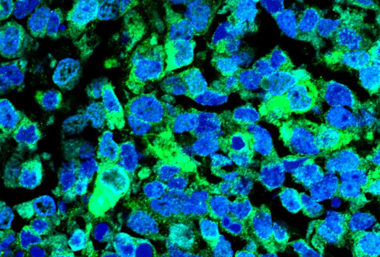
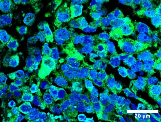
“Ewing sarcoma occurs when two genes are accidentally fused, creating an abnormal protein,” said Vlassakis, a Cancer Prevention and Research Institute of Texas Scholar. “This single change makes cancer cells very good at growing quickly and spreading to other parts of the body, but scientists do not fully understand why.”
The research has a two-part goal. First, Vlassakis aims to develop a test that can determine which proteins bind DNA as well as the abnormal protein that is a signature of Ewing sarcoma. These proteins are likely involved in making some tumor cells better at dividing or spreading.
Second, Vlassakis will use advanced microscopy techniques to visualize how DNA molecules, which in human cells can reach lengths of about 6 feet long, are folded inside cells. The three-dimensional structure that DNA molecules form when folded up determines which genes are active. However, seeing all their kinks and loops in detail is challenging because certain proteins can obscure the view.
“We will use an electric field to remove proteins that hinder our ability to see these tiny DNA structures, each of which is as small as about one thousandth the width of a human hair strand,” Vlassakis said.
By developing new tools to understand how specific genes are regulated in Ewing sarcoma at the cellular level, the work could enable researchers and clinicians to distinguish between different types of cancer cells within the same tumor ⎯ not only for Ewing sarcoma, but potentially for other cancers as well.
“We are fundamentally motivated by a vision of the future of pediatric, adolescent and young adult cancer care in which treatments are personalized, targeting specific molecules to maximize survival and improve the quality of life of survivors,” Vlassakis said.