
Rice University scientists have developed the first complete, label-free molecular atlas of the Alzheimer’s brain in an animal model. The findings help advance understanding of Alzheimer’s onset and progression, a disease that kills more people than breast cancer and prostate cancer combined.
The Rice team used a light-based imaging technique and machine learning to analyze tissue samples from both healthy and diseased brains. The findings, published in ACS Applied Materials and Interfaces, show that Alzheimer’s-related chemical changes are unevenly distributed across the brain and extend beyond the disease’s most familiar hallmark, amyloid plaques.
To capture those changes, the researchers relied on hyperspectral Raman imaging, an advanced form of Raman spectroscopy that uses a laser to read the chemical signatures of molecules.
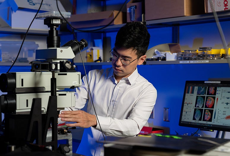
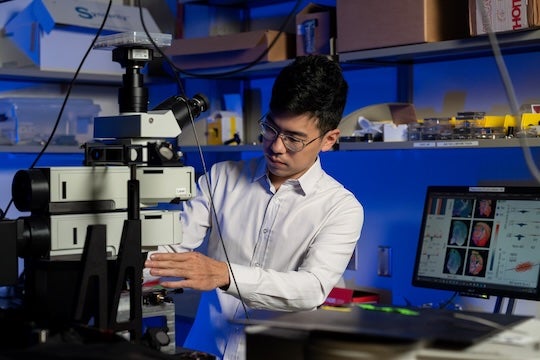
“Traditional Raman spectroscopy takes one measurement of chemical information per molecular site,” said Ziyang Wang, an electrical and computer engineering doctoral student at Rice who is a first author on the study. “Hyperspectral Raman imaging repeats this measurement thousands of times across an entire tissue slice to build a full map. The result is a detailed picture showing how chemical composition varies across different regions of the brain.”
The researchers used the technique to map whole brains, tissue slice by tissue slice, generating detailed molecular maps of healthy and diseased brains from thousands of overlapping spectra. The imaging data obtained was label free, meaning the analyzed samples had not been treated with any dyes, fluorescent proteins or molecular tags.
“This means we observed the brain as is, capturing a complete, unaltered portrait of its chemical makeup,” Wang said. “I think this makes the approach more unbiased and better suited for discovering new disease-related changes that might otherwise be missed.”
To make sense of the vast amount of data collected, the researchers turned to machine learning (ML). They first applied unsupervised ML, which identifies patterns without being told what to look for ahead of time. These models classified tissue based solely on the characteristics of its molecular signals. Next, the team used supervised ML, training models on known Alzheimer’s and non-Alzheimer’s samples. The outputs revealed how strongly different regions of the brain reflect Alzheimer’s-related chemistry.

“We found that the changes caused by Alzheimer’s disease are not spread evenly across the brain,” Wang said. “Some regions show strong chemical changes, while others are less affected. This uneven pattern helps explain why symptoms appear gradually and why treatments that focus on only one problem have had limited success.”
The analysis also uncovered broader metabolic differences between healthy and diseased brains. Cholesterol and glycogen levels varied across disparate brain regions with the starkest contrast found in areas linked to memory ⎯ especially in the hippocampus and cortex.
“Cholesterol is important for maintaining brain cell structure, and glycogen serves as a local energy reserve,” said Shengxi Huang, associate professor of electrical and computer engineering and materials science and nanoengineering and corresponding author on the study. “Together, these findings support the idea that Alzheimer’s involves broader disruptions in brain structure and energy balance, not only protein buildup and misfolding,” added Huang, who is also a member of the Ken Kennedy Institute, the Rice Advanced Materials Institute and the Smalley-Curl Institute.
The idea for the project germinated from long discussions about how to study the Alzheimer’s brain in a different way.
“At first, we were measuring only small areas of brain tissue,” Wang said. “Then I thought, what if we could map the entire brain and gain a much broader view? It took several rounds of testing and trial and error before the measurements and analysis worked well together.”
Wang remembers the excitement of seeing the full chemical map of the Alzheimer’s brain for the first time.
“Patterns emerged that had not been visible under regular imaging,” Wang said. “Seeing those results was deeply satisfying. It felt like revealing a hidden layer of information that had been there all along, waiting for the right way to be analyzed.”
By providing the first detailed chemical maps of the Alzheimer’s brain created without dyes or other molecular markers, the study unlocks a new perspective on the disease as a whole. The researchers say they hope that building on this knowledge could enable earlier detection and better strategies to slow disease progression.
The research was supported by the National Science Foundation (2246564, 1934977), the National Institutes of Health (1R01AG077016) and the Welch Foundation (C2144). The content in this press release is solely the responsibility of the authors and does not necessarily represent the official views of funding entities.
- Peer-reviewed paper:
-
Machine Learning-Enhanced Hyperspectral Raman Imaging for Label-Free Molecular Atlas of Alzheimer’s Brain | ACS Applied Materials and Interfaces | DOI: 10.1021/acsami.5c22623
Authors: Ziyang Wang, Jeewan Ranasinghe, Dennis Chan, Ashley Gomm, Rudolph Tanzi, Can Zhang, Nanyin Zhang and Shengxi Huang
https://doi.org/10.1021/acsami.5c22623 - Access associated media files:
-
Photos:
https://rice.app.box.com/s/6gn1kseivfra6cr3f9mgdtlmxrtby374
(Credit: Jorge Vidal/Rice University)
Video:https://www.youtube.com/watch?v=gNn65aVIs9o
(Credit: Jorge Vidal/Rice University)