

An electrical fingerprint found on tiny particles in the blood may help detect signs of pancreatic cancer that are often overlooked, according to new research from Rice University. The researchers developed a device that separates these particles based on their electrical charge, making cancer-associated signals more visible.

This innovative approach could provide a new method for studying a cancer that is frequently diagnosed after it has already spread with a five-year survival rate of only 13%. The findings were published Sept. 1 in the journal ACS Nano.
“The bloodstream is an incredibly crowded environment, so the challenge is not simply finding tiny particles but identifying the ones that carry meaningful information,” said Kshipra Kapoor, the study’s co-lead author and a Rice electrical and computer engineering doctoral alumna. “Our results show that electrical charge gives us another way to sort through that noise and bring a cancer-associated signal into view.”
Identifying an electrical fingerprint
Cells release tiny packages called extracellular vesicles (EVs) into the bloodstream. These particles can contain material from the cells that created them, including cancer cells. However, finding tumor-derived EVs in blood can be like looking for a needle in a haystack, Kapoor said, because they are mixed among a much larger population of EVs released by healthy tissues.
The researchers, including Rice PhD candidate Paul Spezza, who contributed equally to this study, aimed to determine whether pancreatic cancer leaves a distinct physical mark on these EVs. They focused on the KRAS gene, which acts as a switch for cell growth. When KRAS is mutated, this switch can become stuck in the “on” position, allowing uncontrolled cell growth. Mutations in KRAS are a major factor in pancreatic cancer.
The research team discovered that activating mutant KRAS caused pancreatic cancer cells to release more EVs, which also exhibited a stronger negative electrical charge. The presence of DNA and other materials on the outer surface contributed to this electrical signature.
“Pancreatic cancer appears to leave an electrical fingerprint on the tiny packages that cells send into the bloodstream,” Kapoor said. “That fingerprint could give us a new way to pick out cancer-related EVs that would otherwise be lost in the crowd.”
Using electricity to isolate cancer signals
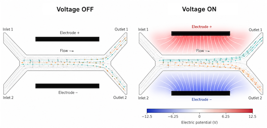
The researchers designed a small device that uses electricity to separate EVs based on their charge. This microfluidic electrophoresis device moves minute amounts of blood serum through narrow channels, allowing for the collection of EVs with a stronger negative charge.
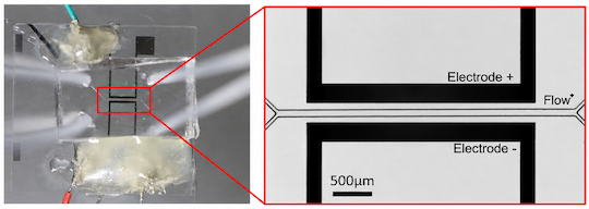
The device provides researchers with a new method to search for cancer-related EVs. Many existing techniques separate these particles based on size or weight or use specially designed molecules for capture, Kapoor said. In contrast, this device capitalizes on a physical trait that EVs already possess: their electrical charge.
The researchers tested the device on 112 samples from healthy individuals, patients with pancreatic cancer and patients with pancreatitis, or inflammation of the pancreas. The goal was to see if the electrical differences observed in lab-grown cells would also be evident in samples from people.
Revealing hidden cancer signals
Simply measuring the average electrical charge of all EVs in the blood proved insufficient for distinguishing pancreatic cancer samples from healthy ones. The cancer-associated particles were effectively masked by the much larger population of vesicles circulating in the bloodstream.
However, once the device separated the vesicles, the differences became clearer. Samples from patients with pancreatic cancer showed greater enrichment of the more negatively charged EV population.
This result suggests that sorting particles based on a physical characteristic can uncover information that an average measurement of the entire population might overlook.
“An electrocardiogram turns the heart’s electrical activity into information doctors can use,” Kapoor said. “In a similar spirit, our work suggests that the electrical properties of extracellular vesicles could one day provide another way to read biological information from cancer.”
Other authors of the study include Rice researchers Paul Spezza, Beatrice Pforr, Martin Bell, Bo Fan, Yi-Lin Chen, Sibani Lisa Biswal, Xin Luo, Kaira Church, Seoyun Kong, Elena Ramirez, Fernanda Kugeratski, Jacob Robinson and Raghu Kalluri; Kathleen McAndrews from The University of Texas MD Anderson Cancer Center; Florian Gebauer from Helios University Hospital Wuppertal; and Christoph Kahlert from Heidelberg University.
The study received support from the National Cancer Institute, the Sid W. Richardson Foundation and MD Anderson.