
The spinal cord is harder to access and study than even the brain. The challenges posed by its mobility and anatomical structure have made understanding exactly how it functions difficult.
Rice University engineers will work with collaborators to optimize an array of nanoelectronic threads, or NETs ⎯ already used successfully for gathering high-fidelity, long-term data from neurons in the brain ⎯ for use in the spine, supported by a $6.25 million, four-year grant from the National Institutes of Health.
In addition to neuronal activity recordings, NET probes can provide tunable, localized stimulation of adjacent neurons. Rice neuroengineers also hope to maximize NETs' functional bandwith by integrating them into a larger-scale, data-processing system.
The new tool could help neuroscientists crack the secrets of spinal cord function and bring new hope to patients dealing with injuries and other associated medical conditions.
“So far, we haven't had a good understanding of how the neurons in the spinal cord actually work,” said Chong Xie, the principal investigator on the grant and an associate professor of electrical and computer engineering and neuroengineering. “For example, if you move your arms or walk around, you have the intention in your brain and the muscles operate exactly as you want them to. This conversion of the initial intention into specific motions of each of the muscles is operated and implemented in the spinal cord, where circuits consisting of many, many neurons are responsible for carrying out this job. But we don't know exactly how this is achieved.”
Using electrodes to track neuronal activity in the brain has allowed neuroscientists to learn a great deal about brain function. The flexible NET probes developed by Xie and collaborators integrate seamlessly with brain tissue and perform better than rigid probes when used to record electrical information from individual neurons in the brain.
Preliminary tests have shown that NET probes can achieve high-quality, long-duration recordings from spinal cord neurons of mice. However, the scientists intend to further adapt NETs to the specific structural and functional demands of the spinal cord.
In the brain, the distribution of neurons, or gray matter, and the bundles of nerve fibers known as white matter is the exact inverse of spinal cord anatomy.
“We typically refer to this as the ‘inside-out anatomy’ of the spinal cord,” said Lan Luan, an assistant professor of electrical and computer engineering and co-investigator on the grant. “The outer layer of the brain ⎯ the gray matter ⎯ is where the neurons are, whereas the fibers called white matter are on the interior. In the spinal cord, the white matter or fibers are on the exterior, shielding the neurons. This makes accessing those neurons more challenging.”
To ensure better access, scientists plan to develop a probe design that is small enough to be implanted at different sites on the spine yet has greater depth coverage and enough channels to capture data from neurons in a spinal cord cross section.
Another goal is to equip the probes with stimulation capabilities in addition to their recording function.
“The electrode can do both,” Luan said. “This has a direct health relevance, because for patients with spinal cord injury or other types of injuries, stimulation could be a way to restore fine motor control. There are several very successful technologies demonstrating that stimulation in the cord can restore local motions. But to impact finer motor control, we believe we need to go inside the cord and have a greater degree of access and precision for applying this stimulation.”
The spinal cord plays a significant role in pain processes, so identifying which spinal neurons are directly involved with pain-signal relay could open the door to better pain-management therapies.
“Identifying the specific type of spinal neurons that play a significant role in processing pain information could potentially enable the development of drugs that target precisely those cells,” Xie said. “Or maybe we can use the electrodes to stimulate those neurons and modulate their activity so that they don’t convey the pain signal to the brain.”
Scientists plan to not only optimize probe design, but also to incorporate spinal NETs into an extremely miniaturized, integrated data-processing and stimulation-feedback system.
In addition to developing the technology, Xie, Luan and their team have partnered with the Pfaff lab at the Salk Institute for Biological Studies, the Weber lab at Carnegie Mellon University and the Basbaum and Ganguly labs at University of California, San Francisco for a series of spinal cord studies that will test the devices across different spinal regions, animal models and research topics.
Luan said she hoped that developing and optimizing NET-based technology for spinal cord research would “provide a tool that can help the entire neuroscience community achieve a more fundamental understanding of spinal cord function.”
“My real hope is that, four years down the road, at the end of this project, neuroscientists will be able to see and do new things that are impossible with current technology,” Xie said.
- Award information:
-
Award number: U01NS131086-01
Title: “Optimizing ultraflexible electrodes and integrated electronics for high-resolution, large-scale intraspinal recording and modulation” - Image downloads:
-
https://news-network.rice.edu/news/files/2023/04/230330_NIH-U01-Grant-Chong-Xie-Lan-Luan_1_LG.jpg
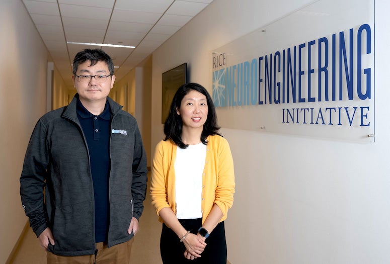
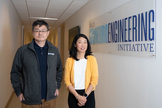
CAPTION: Chong Xie (left) is the principal investigator on the grant and an associate professor of electrical and computer engineering and neuroengineering at Rice. Lan Luan is an assistant professor of electrical and computer engineering and co-investigator on the grant. (Photo by Gustavo Raskosky/Rice University)
- Related stories:
-
Engineers explore the source of dreams and thoughts:
https://news.rice.edu/news/2022/engineers-explore-source-dreams-and-thoughts
Gentle probes could enable massive brain data collection:
https://news.rice.edu/news/2020/gentle-probes-could-enable-massive-brain-data-collection
Blood flow recovers faster than brain in micro strokes:
https://news.rice.edu/news/2020/blood-flow-recovers-faster-brain-micro-strokes - Links:
-
Luan lab: https://www.luanlab.net/
Xie lab: https://www.chongxie.net/
Rice Neuroengineering Initiative: https://neuroengineering.rice.edu/
Rice Integrated Systems and Electromagnetics Lab (Chi group): https://chilab.info
Department of Electrical and Computer Engineering: https://eceweb.rice.edu
- About Rice:
-
Located on a 300-acre forested campus in Houston, Rice University is consistently ranked among the nation’s top 20 universities by U.S. News & World Report. Rice has highly respected schools of Architecture, Business, Continuing Studies, Engineering, Humanities, Music, Natural Sciences and Social Sciences and is home to the Baker Institute for Public Policy. With 4,552 undergraduates and 3,998 graduate students, Rice’s undergraduate student-to-faculty ratio is just under 6-to-1. Its residential college system builds close-knit communities and lifelong friendships, just one reason why Rice is ranked No. 1 for lots of race/class interaction and No. 4 for quality of life by the Princeton Review. Rice is also rated as a best value among private universities by Kiplinger’s Personal Finance.