Biochemist George Phillips ’74 is back for a third stint at Rice
George Phillips ’74 is both a new face on campus and a 20-year Rice veteran, and if that is puzzling, it’s also poetic because Phillips’ quest to solve biological puzzles began at Rice almost 40 years ago and brought him back for good last fall.
Phillips, the Ralph and Dorothy Looney Professor of Biochemistry and Cell Biology and professor of chemistry, first arrived at Rice as an undergraduate in 1970 and left six years later with two bachelor’s degrees (chemistry and biochemistry) and Rice’s first Ph.D. in biochemistry. He returned twice as a tenured professor, first in 1987 and most recently after a 12-year stint at the University of Wisconsin, Madison.
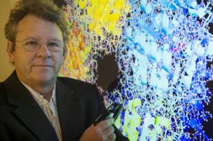
Phillips in the 3-D visualization studio he recently constructed in his group's conference room.
Phillips’ love of crystallography and structural biology began at Rice and fueled his career. His lab has deciphered the structures of 363 proteins, and his published research runs the gamut from diffraction physics to computer science. Phillips also had a hand in starting the Gulf Coast Consortium, the organization that spearheads more than a dozen multi-institutional partnerships in the Texas Medical Center.
Rice News caught up with Phillips this month as he was putting the finishing touches on a 3-D visualization studio he’d just constructed in his lab’s conference room using a commercially available 80-inch 3-D television and off-the-shelf computer software.
Tell us about your research.
I am essentially in the molecular architecture business. We also study protein engineering and structure-function relationships. Just last week, I figured out how to use a standard 3-D TV to project images of the molecular structures we work with. When we solve a new structure, we can come in here and sit and talk about the results. We do this with the help of a little broadcast-company electronics and a standard PC.
Did you write the software yourself?
This particular application already existed, but we do write our own software. When I was at Wisconsin, I had a joint appointment in computer science, and I have published papers in computer science journals on the technology to support crystallography. We have a recent paper — I haven’t got it implemented in here yet — where we developed a technology to use Xbox controllers for people to interact with the objects. Everybody’s got their own cursor, and they can move the molecule around.
For those group meetings?
Yes. Four people can sit at the group meetings — each with their own controller — and say, “Look over here. What about this?” And they can use the thumb joystick to rotate the image around.
What are we looking at?
This is a cellulase. Cellulases are enzymes that break down cellulose. This one is part of a biofuels project that I’m involved in that’s related to cellulosic ethanol production. Cellulase is very hard to break down, and we’re trying to develop better enzymes to do that.
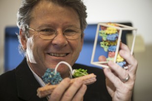
Phillips lab has deciphered the structure of more than 360 proteins, and dozens of these are on display in the form of colorful clay models in his office.
Why biochemistry? What got you into that?
I came to Rice as a physics major, but I realized there were many students who were much better at physics than I was. There was an open house over in the Chemistry Department, and Ron Sass was a crystallographer in the Chemistry Department at that time. He had a bunch of molecular models in the lab, and people were sitting around looking at these odd printouts and objects. It was explained to me by one of the researchers there that crystallography was kind of like solving puzzles. I’d always liked puzzles, and I said, “Wait a minute. You can get paid to solve puzzles?
Soon after that, Florante Quiocho, a young assistant professor who later became my Ph.D. adviser, put up this structure in class of a protein molecule in all its atomic glory. He said the protein’s structure had been solved by crystallography. I thought, “Wow, there are thousands of atoms, and you can figure out where they all are, look at the molecular architecture and explain how they work.” I was hooked at that point.
What do puzzles and X-ray crystallography have in common?
To image these molecules with X-ray crystallography, you first grow a crystal that has a whole bunch of the individual protein molecules all lined up in a regular pattern, like little soldiers. When you shine X-rays on the crystal, you get constructive and destructive interference that leads to a diffraction pattern, and the puzzle is how you go backward from that diffraction pattern to the arrangement of atoms that gave rise to that pattern. In optimization theory, that’s called an inverse problem. You don’t care that much about the diffraction pattern; you care about inverting it to give the location of all the atoms. And that’s a puzzle because you don’t really have all of the information you need. When we solve a new structure, that means we’ve solved this inverse problem and we’ve gotten back to the location of the atoms.
Is it also a puzzle to decipher the function of the protein or the ways that it interacts with other molecules? Are these puzzles within puzzles?
Yes. How does it work? And how does the structure relate to what it does? Those are puzzles also. In structural biology we are ultimately interested in the structure-function relationships — the connection of three-dimensional molecular structures to the function they perform in the organism.
There are other ways to get structure other than crystallography. Structural biologists can also use nuclear magnetic resonance and mass spectrometry to some degree. They can also use electron microscopy.
But crystallography was first, right? It’s been around a long time.
William Bragg and his son, Lawrence Bragg, got the Nobel Prize in 1915 for showing you could use crystallography to solve the structure of sodium chloride. In fact, 2014 has been declared by the United Nations as the International Year of Crystallography in celebration of the work of the Bragg father-son team and Max Von Laue, a German scientist who got the Nobel Prize the year before they did for his work on explaining X-ray diffraction.
How far has the field progressed in the last century?
Some of the cool things happening in crystallography right now are the development of new X-ray sources. There’s an X-ray laser out at Stanford that I’m doing some research with. Another area is the development of high-throughput crystallization methods, which take advantage of robotic imaging and robotic production of combinatorial solutions for creating crystals. When I was a student, if you solved a structure over five years, you really had something. Now, there was a period when my lab solved about 100 structures in one year. We’re not in that mode any more, but the tools for making crystallography routine are now quite common and quite powerful.
The latest tools allow you to work faster and go after more difficult proteins. One example would be membrane proteins. Those are proteins that are embedded in membranes as opposed to being soluble proteins. About a third of all proteins are membrane proteins. We know the structure of more than 50,000 soluble proteins, but we have structures for only a few hundred membrane proteins.


Leave a Reply